
Makassar, Indonesia Aug 1 2026/ADDIS/ – Modern lifestyle shifts toward high-calorie diets rich in saturated fats and cholesterol have significantly increased metabolic health concerns worldwide. Excessive intake of dietary fat contributes to dangerous lipid accumulation across vital internal organs, raising the long-term risk of severe cardiovascular conditions, metabolic disorders, and chronic organ damage. Because these pathological shifts often begin silently at the cellular level, early detection is critical for preventing irreversible structural changes. To better understand how high-cholesterol consumption alters internal organ structures in real time, biomedical researchers frequently rely on preclinical animal models to observe subtle structural shifts long before overt clinical symptoms appear.
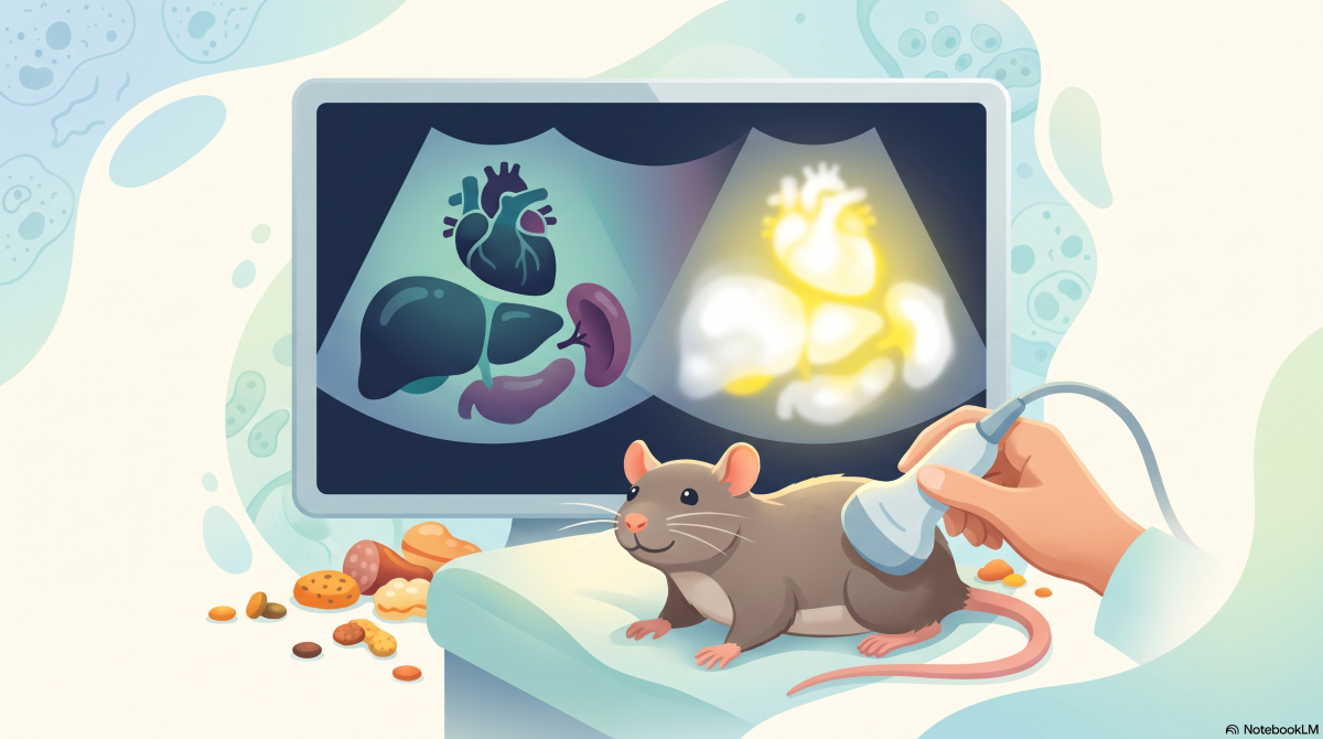
A biomedical research study published on January 12, 2025, in the International Journal of Veterinary Science by Unique Scientific Publisher, titled “Abdominal Ultrasonography Imaging after High-Fat Diet Administration in Wistar Mice (Rattus norvegicus)”, investigated the visual impact of high-fat dietary intake on vital internal body structures. The research was conducted by scientists affiliated with the Infectious Animal Disease Dynamics Thematic Research Group (ADDIS TRG). The team evaluated male Wistar laboratory rats (R. norvegicus) subjected to varying proportions of high-fat diets over a 30-day experimental period. By utilizing non-invasive abdominal ultrasonography (USG), the study aimed to observe structural changes and fat deposition patterns across three key internal organs: the heart, the liver, and the spleen.
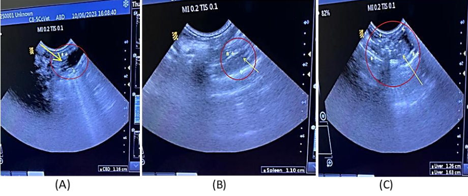
The ultrasound imaging revealed stark visual differences between healthy control animals and those receiving high-fat diets. While control groups exhibited well-defined organ boundaries and normal tissue density, all dietary treatment groups (ranging from 0.5% to 2% fat intake relative to total body weight) displayed abnormal hyperechoic—or bright white—reflections across the heart, liver, and spleen. In the heart, accumulated fat obscured the internal boundaries between the atrial and ventricular cardiac cavities. Similarly, the liver and spleen displayed diffuse hyperechoic changes across their margins, indicating widespread lipid accumulation and tissue disruption.
These ultrasound findings carry profound relevance for preventive medicine. Non-invasive ultrasonography provides a clear visual baseline for monitoring metabolic disease progression without requiring invasive surgical procedures. Identifying early structural changes in organ tissue helps researchers evaluate the systemic consequences of lipid accumulation, supporting human metabolic health studies while simultaneously enhancing clinical veterinary care for companion animals facing obesity-related conditions.
This research initiative directly supports the United Nations Sustainable Development Goal (SDG) 3: Good Health and Well-Being. By advancing early diagnostic techniques for lipid-induced organ damage, the study reinforces broader global efforts to understand, prevent, and manage metabolic and cardiovascular health challenges across species.
In conclusion, abdominal ultrasonography serves as an effective, accessible diagnostic tool for detecting dietary-induced fat accumulation in internal organs. Establishing clear imaging markers for lipid deposition highlights the vital necessity of balanced nutrition in preventing metabolic disease, offering valuable insights for both veterinary diagnostics and human health research.
DOI:
https://doi.org/10.47278/journal.ijvs/2025.003
Contact person:
Prof. Dwi Kesuma Sari
+62 852-9927-9988
dwiksari@vet.unhas.ac.id