Shell Deformity in the Green Mussel (Perna viridis): A Candidate Morphological Biomarker
Shell Deformity in the Green Mussel (Perna viridis): A Candidate Morphological Biomarker
Khusnul Yaqin
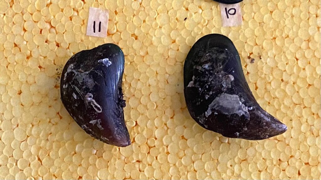
The green mussel (Perna viridis) has long been recognized as an effective bioindicator for assessing environmental quality in coastal waters. One of the most commonly observed phenomena in recent years is shell deformity—morphological irregularities characterized by asymmetry, abnormal curvature, and uneven shell growth (Yaqin, 2004; Yaqin, 2022). A recent study by Yap et al. (2023) reported shell deformity prevalence as high as 87.5% along the west coast of Peninsular Malaysia—an alarming rise compared to previous reports of 0–36.8%. This trend is strongly associated with increasing exposure to metals such as cadmium (Cd), lead (Pb), copper (Cu), and zinc (Zn), which have been detected at elevated levels in the soft tissues and shells of P. viridis.
Shell formation in bivalves is governed by calcium carbonate (CaCO₃) biomineralization, a complex process regulated by matrix proteins, enzymes, and signalling molecules. Among the most critical regulators of this process is calmodulin (CaM), a highly conserved calcium-binding protein present in all eukaryotic cells. Calmodulin functions as a calcium sensor, activating specific enzymes and modulating the expression of proteins essential for shell matrix construction. Under normal physiological conditions, calmodulin binds Ca²⁺ and facilitates the orderly deposition of aragonite or calcite crystals that compose the shell layers. Studies on other bivalves such as Pinctada fucata and Crassostrea gigas have demonstrated that calmodulin-like proteins (CaLPs) are actively expressed during periods of shell growth and regeneration (Yan et al., 2007; Lin et al., 2021).
However, toxic metals such as Cd and Pb can disrupt calmodulin function by substituting for Ca²⁺ at EF-hand binding domains. This substitution alters calmodulin’s conformation, impairing its ability to initiate and regulate the biomineralization pathways. Consequently, the crystalline architecture of calcium carbonate becomes disorganized, resulting in visible shell deformities. Proteomic analyses by Li et al. (2005) confirmed that calmodulin-like proteins are integral to aragonite crystal formation, and that interference by toxic metals leads to structural degradation and diminished protein activity. Subsequent studies have shown that calmodulin gene expression is highly sensitive to chemical stress. Metal-induced mutations or transcriptional disruptions reduce internal Ca²⁺ homeostasis, compromising the biological machinery necessary for proper shell synthesis (Xia et al., 2018; Li et al., 2020).
Thus, impairment of calmodulin function has consequences that extend from the molecular to the morphological level. Shell deformities resulting from calmodulin disruption are not only physiologically meaningful but also visibly apparent, making them powerful indicators of chemical stress in marine environments. In this context, shell deformities in P. viridis may serve as a cost-effective, field-applicable morphological biomarker of metal contamination. To ensure scientific validity, however, this approach requires an integrative framework—combining morphometric analysis with molecular assessments of calmodulin expression, quantification of metal accumulation in tissues and shells, and environmental parameters such as pH, dissolved oxygen, and salinity.
The development of shell deformity as a morphological biomarker underpinned by calmodulin-based molecular mechanisms represents a novel avenue in coastal ecotoxicology. This direction not only enhances our understanding of metal toxicity at the cellular level but also provides an actionable tool for environmental monitoring, enabling more informed and evidence-based decision-making in marine conservation and coastal management.
References
- Li, S. et al. (2005). “cDNA cloning and characterization of a novel calmodulin‐like protein from pearl oyster Pinctada fucata.” The FEBS Journal, 272(19), 4899–4910.
- Li, Y. et al. (2020). “Molecular responses of an estuarine oyster to multiple metal contamination in Southern China revealed by RNA-seq.” Science of the Total Environment, 701, 134648.
- Xia, X. et al. (2018). “Characterization of calmodulin in the clam Anodonta woodiana: differential expressions in response to environmental Ca²⁺ and Cd²⁺.” Turkish Journal of Biochemistry, 43(4), 403–416.
- Yap, C. K. et al. (2023). “Shell Deformities in the Green-Lipped Mussel Perna viridis: Occurrence and Potential Environmental Stresses on the West Coast of Peninsular Malaysia.” Pollutants, 3(3), 406–418.
- Yaqin, K. (2004). “Ada Kerang Abnormal di Teluk Jakarta.” Kompas, p. 4.
- Yaqin, K. (2022). “DNA Damage and Shell Malformation in Blue Mussel, Mytilus edulis.” Akuatikisle: Jurnal Akuakultur, Pesisir dan Pulau-Pulau Kecil, 6(1), 65–74.